Phone: 813.792.4804
Diagnostic Ultrasound for Muscular/Skeletal Diseases
 Ultrasound imaging, also called ultrasound scanning or sonography, involves exposing part of the body to high-frequency sound waves to produce pictures of the inside of the body. Ultrasound exams do not use ionizing radiation (as used in x-rays). Because ultrasound images are captured in real-time, they can show the structure and movement of the body's internal organs, as well as blood flowing through blood vessels.
Ultrasound imaging is a noninvasive medical test that helps physicians diagnose and treat medical conditions.
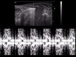
Ultrasound images of the musculoskeletal system provide pictures of muscles, tendons, ligaments, joints and soft tissue throughout the body.
What are some common uses of the procedure?
Ultrasound images are typically used to help diagnose:
- tendon tears, such as tears of the rotator cuff in the shoulder or Achilles tendon in the ankle.
- abnormalities of the muscles, such as tears and soft-tissue masses.
- bleeding or other fluid collections within the muscles, bursae and joints.
- small benign and malignant soft tissue tumors.
- early changes of rheumatoid arthritis.
- Ultrasound images can also be used for fractures.
How does the procedure work?
Ultrasound imaging is based on the same principles involved in the sonar used by bats, ships and fishermen. When a sound wave strikes an object, it bounces back, or echoes. By measuring these echo waves it is possible to determine how far away the object is and its size, shape, and consistency (whether the object is solid, filled with fluid, or both).
In medicine, ultrasound is used to detect changes in appearance of organs, tissues, and vessels or detect abnormal masses, such as tumors.
In an ultrasound examination, a transducer both sends the sound waves and records the echoing waves. When the transducer is pressed against the skin, it directs small pulses of inaudible, high-frequency sound waves into the body. As the sound waves bounce off of internal organs, fluids and tissues, the sensitive microphone in the transducer records tiny changes in the sound's pitch and direction. These signature waves are instantly measured and displayed by a computer, which in turn creates a real-time picture on the monitor. One or more frames of the moving pictures are typically captured as still images.
What will I experience during and after the procedure?
Most ultrasound examinations are painless, fast and easy. After you are positioned on the examination table, the radiologist or sonographer will apply some warm water-based gel on your skin and then place the transducer firmly against your body, moving it back and forth over the area of interest until the desired images are captured. There is usually no discomfort from pressure as the transducer is pressed against the area being examined.
If scanning is performed over an area of tenderness, you may feel pressure or minor pain from the transducer.
The radiologist or sonographer may ask you to move the extremity being examined or may move it for you to evaluate not only anatomy but also function of a joint, muscle, ligament or tendon.
Once the imaging is complete, the gel will be wiped off your skin. After an ultrasound exam, you should be able to resume your normal activities within a few hours.
What are the benefits?
Benefits
- Most ultrasound scanning is noninvasive (no needles or injections) and is usually painless.
- Ultrasound is widely available, easy-to-use and less expensive than other imaging methods.
- Ultrasound imaging uses no ionizing radiation.
- Ultrasound scanning gives a clear picture of soft tissues that do not show up well on x-ray images.
- Ultrasound causes no health problems and may be repeated as often as is necessary.
- Ultrasound provides real-time imaging, making it a good tool for guiding minimally invasive procedures such as needle biopsies and needle aspiration.
- Unlike the strong magnetic field of magnetic resonance imaging (MRI), ultrasound is not affected by cardiac pacemakers, ferromagnetic implants or fragments within the body. Ultrasound is also an excellent alternative to MRI for claustrophobic patients.
- Ultrasound may actually have advantages over MRI in seeing tendon structure, which is better appreciated by ultrasound than MRI.
If you want to find out more Contact Us today for consultation.